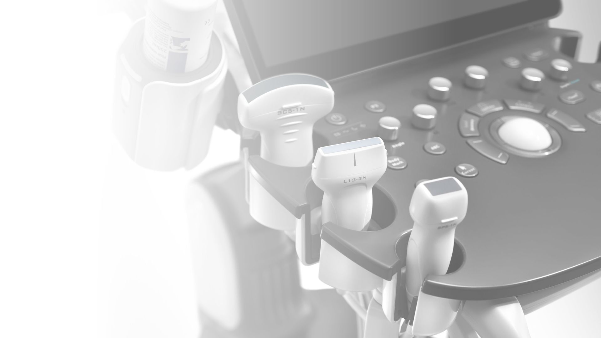
Ultrasonography
Linove doctors' practice offers a wide range of ultrasonographic examinations:
- Ultrasonography of joints, muscles and ligaments
- Ultrasonography of thyroid and neck soft tissues
- Ultrasonography of organs of the abdominal cavity
- Dopplerography of the vessels of the neck and head
- Dopplerography of leg veins and arteries
Ultrasonography is one of the most frequently used diagnostic methods chosen by specialists to clarify a number of diagnoses. It is used in cases of tendon, ligament or muscle injuries to specify the severity of the damage. Ultrasonographic examinations of the organs of the abdominal cavity are an integral part of the diagnostics of the digestive organ system. With the help of ultrasonography, it is possible to evaluate the condition of the liver, gallbladder and bile ducts, spleen, kidneys, pancreas, bladder, as well as the flow of blood vessels in the abdominal cavity. Ultrasonography is also often used to evaluate the state of the thyroid gland.
Ultrasonography is a safe and affordable examination method for the patient
Musculoskeletal or ultrasonography of joints, ligaments, tendons and muscles.
Ultrasonography is ideal for diagnosing superficial organs. Most of our body's ligaments, tendons, and muscles lie superficially, making them a good target for ultrasound examinations. Thanks to the fact that the examination can be performed during active and passive movement of the body, it is possible to evaluate the organs of the support and movement system in dynamics, which allows to see the pathological changes that cannot be diagnosed in a neutral state.
By examining the joints, ligaments, tendons and muscles, it is possible to diagnose such common problems as:
- Osteoarthritis of the joints
- Inflammation of the joints
- Tearing of tendons, ligaments or muscles
- Inflammation of tendons, ligaments or muscles
- Bursitis
- Calcifying tendinitis
- Benign and malignant formations
Under ultrasonography control, it is possible to purposefully administer preparations that improve the effectiveness of injections and reduce the risks associated with joint or periarticular injections. Under the control of ultrasonography, it is possible to perform:
- Injections of anti-inflammatory drugs in and around joints, around tendons, ligaments and muscles
- Platelet rich plasma (PRP) injections into joints, peri-articular, around tendons, ligaments and muscles
- Carry out treatment of calcifying tendinitis using barbotage procedure
- Perform other types of therapeutic and diagnostic invasive procedures
Ultrasonography of organs of the abdominal cavity.
Abdominal ultrasonography is one of the most requested ultrasonographic examination methods. Ultrasonography of abdominal cavity organs is most often used for the diagnosis of pathology of organs of the digestive tract, kidneys and organs of the urinary system. The examination has several advantages, the main one being safety. Ultrasonography of abdominal organs does not cause side effects and is safe for pregnant women and children.
By examining the organs of the abdominal cavity, it is possible to diagnose the following common problems:
- Hepatic steatosis (fatty liver)
- Bile reflux disorder
- Gallstones
- Kidney stones
- Impaired bladder emptying
- Inflammation of the organs of the abdominal cavity
- Benign and malignant formations
Ultrasonography of thyroid and neck soft tissues.
The thyroid gland is a superficial organ that is ideally suited for diagnosis by ultrasonography. Thyroid nodules are a common pathology among the population of Latvia. Ultrasonography helps to determine several signs that allow the doctor to understand whether the formation is malignant and, if necessary, order a biopsy of the thyroid nodule, which allows to determine the nature of the nodule with high reliability.
During the examination, the doctor examines not only the tissues of the thyroid gland, but also the surrounding tissues where several lymph nodes are located, and also evaluates the salivary glands and other structures in the soft tissues of the neck.
Doppler imaging of blood vessels.
With the help of ultrasonography, it is possible to perform a doppler examination. When examining the blood vessels, the doctor can determine the blood flow rates in the lumen of the blood vessels, visually evaluate the walls of the blood vessels, determine atherosclerotic changes in the arteries, venous return disorders in the veins. In cases where the arteries are partially closed by atherosclerotic thrombus, the doctor can evaluate the flow rate at the thrombus site and the section of the whole blood vessel, by comparing the flow velocities conclusions can be made about the degree of narrowing of the blood vessel. The greater the acceleration of the flow, the greater the narrowing of the artery.
Frequently asked Questions:
Yes. Ultrasonography is a safe examination method. It is allowed for pregnant women and children. The essence of the investigation method is the reception of ultrasound radiation and its echo, which is not harmful to the human body.
After the procedure, the doctor will immediately create an extract with the results of the examination, explaining its nature.
Before the ultrasonography of the abdominal organs, it is preferable to arrive in an empty shower (not at least 4 hours before the procedure) and with a full bladder. It is also necessary to come to the ultrasonographic examinations of the urinary organs with a full bladder. No special preparation is required for other ultrasonographic examinations.
Ultrasonography is performed by

Dr. Viktors Linovs
Radiologist