Mole Screening and Digital Mapping Using Canfield and DermaGraphix®

Mole screening and digital mapping in a single visit, including a consultation with a dermatologist. The doctor evaluates skin lesions, performs an in-depth examination with a dermatoscope for those who need it, and provides recommendations. Images stored in the Canfield system allow for comparison of skin condition during follow-up examinations and help identify new or changing lesions.

When is it particularly worth considering digital mapping?

  • If you have many moles and find it difficult to keep track of changes in them.
  • If you’ve been diagnosed with melanoma before.
  • If there is a family history of melanoma or a known genetic predisposition.
  • If your dermatologist recommends regularly documenting and comparing skin lesions.


A dermatologist determines the need for monitoring and the frequency of checkups on a case-by-case basis.


Book now

Genital Wart Screening (80 EUR) + Dermatologist Consultation (70 EUR)

+37124401084

Starting at

€ 150,00

In Brief

A Brief Overview of Birthmark Mapping

Before the visit

Before the visit

  • Before the visitPreparing for your visit is simple—please follow the recommendations below before your appointment.
  • Before the visitOn the day of your visit, do not apply makeup or oily or tinted body products to the area of skin being examined.
  • Before the visitAvoid self-tanning products before the scan, as they can make it difficult to evaluate skin lesions and compare images.
  • Before the visitIf you have long hair, bring a hair tie: the neck and scalp must be visible.
  • Before the visitThe photos are taken while you are in your underwear; you and one specialist will be in the room.
  • Before the visitBefore the examination, inform the specialist about any skin lesions, inflammation, or known contact allergies. The doctor will select the appropriate examination method for the specific area.
  • Before the visitThe images will be added to your medical record. Only medical staff will have access to them.
What happens during the procedure

What happens during the procedure

  • What happens during the procedureYou stand on the positioning mat, facing the device.
  • What happens during the procedureA specialist will help you assume the poses required for the imaging. The Canfield IntelliStudio system will capture standardized full-body images, which will be saved for future comparison.
  • What happens during the procedureThe doctor will also examine areas that are not clearly visible in the full-body images, such as the scalp, palms, and soles of the feet. Depending on the findings, additional dermatoscopy and photography will be performed.
  • What happens during the procedureThe device does not touch you—it only takes photos. The dermatoscope touches the skin only in the areas the doctor wants to examine in detail.
  • What happens during the procedureThe visit, including digital mapping, mole screening, and a consultation with a dermatologist, lasts approximately 40–50 minutes.
Results

Results

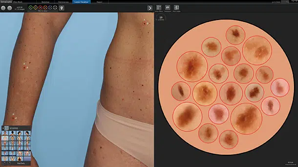
  • ResultsA digital skin map is created, containing full-body overview images and dermatoscopic images of lesions selected by the physician. The documented lesions are assigned identification numbers so they can be located and compared during future visits.
  • ResultsDuring the visit, the dermatologist explains the examination results and provides recommendations: which lesions should be monitored, whether additional testing or removal is necessary, and when the next checkup should take place.
  • ResultsThe dermatologist evaluates the skin lesions and determines the next course of action. Digital images supplement the doctor’s physical examination and dermatoscopy, helping to document the findings and track changes over time.
Frequency

Frequency

  • Frequency

    The greatest value of digital mapping of moles becomes apparent over the long term. Repeated examinations allow for an objective comparison of moles, the detection of even very minor changes, and the early detection of suspicious lesions, including signs of early-stage melanoma.

  • Frequency

    The dermatologist determines the timing of the next examination based on the location of the mole and the individual’s risk. A follow-up may be scheduled in 6–12 months or sooner. The doctor may recommend dermatoscopic monitoring of a specific lesion at shorter intervals; this does not always mean a repeat full-body examination.

  • Frequency

    Don’t forget to check your own skin! Examine your skin once a month and pay attention to any new or changing moles. If you notice any changes, don’t wait for your next scheduled appointment—make an appointment with a dermatologist sooner.

1 /

How does Canfield DermaGraphix® work?

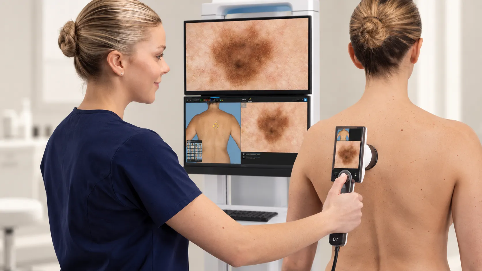
Canfield IntelliStudio provides standardized body photography, while the DermaGraphix software helps organize and compare follow-up and dermatoscopic images. Consistent imaging conditions help the doctor more accurately assess changes between visits.

Full-Body Scan

Full-Body Scan

There is a positioning mat on the floor, and the device projects a thin line of light onto it—it always remains at the same distance. The camera automatically detects the height of your face and moves through four fixed positions.

Comparison of Two Images

Comparison of Two Images

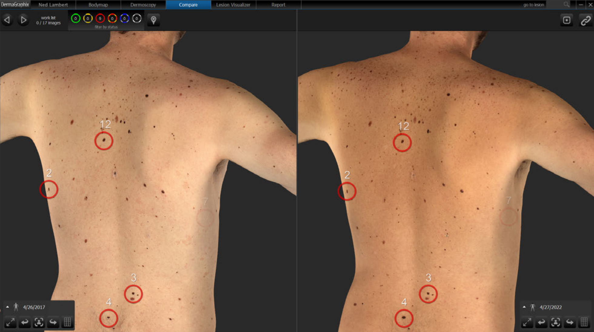
A person never stands in exactly the same position as they did last year. The software helps align images from different visits and compare the same areas of skin. The doctor assesses whether the visible differences indicate new or changing skin lesions. There’s no need to mark the skin or apply stickers.

Dermatoscope — close-up

Dermatoscope — close-up

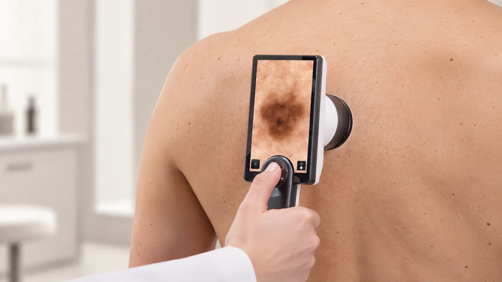
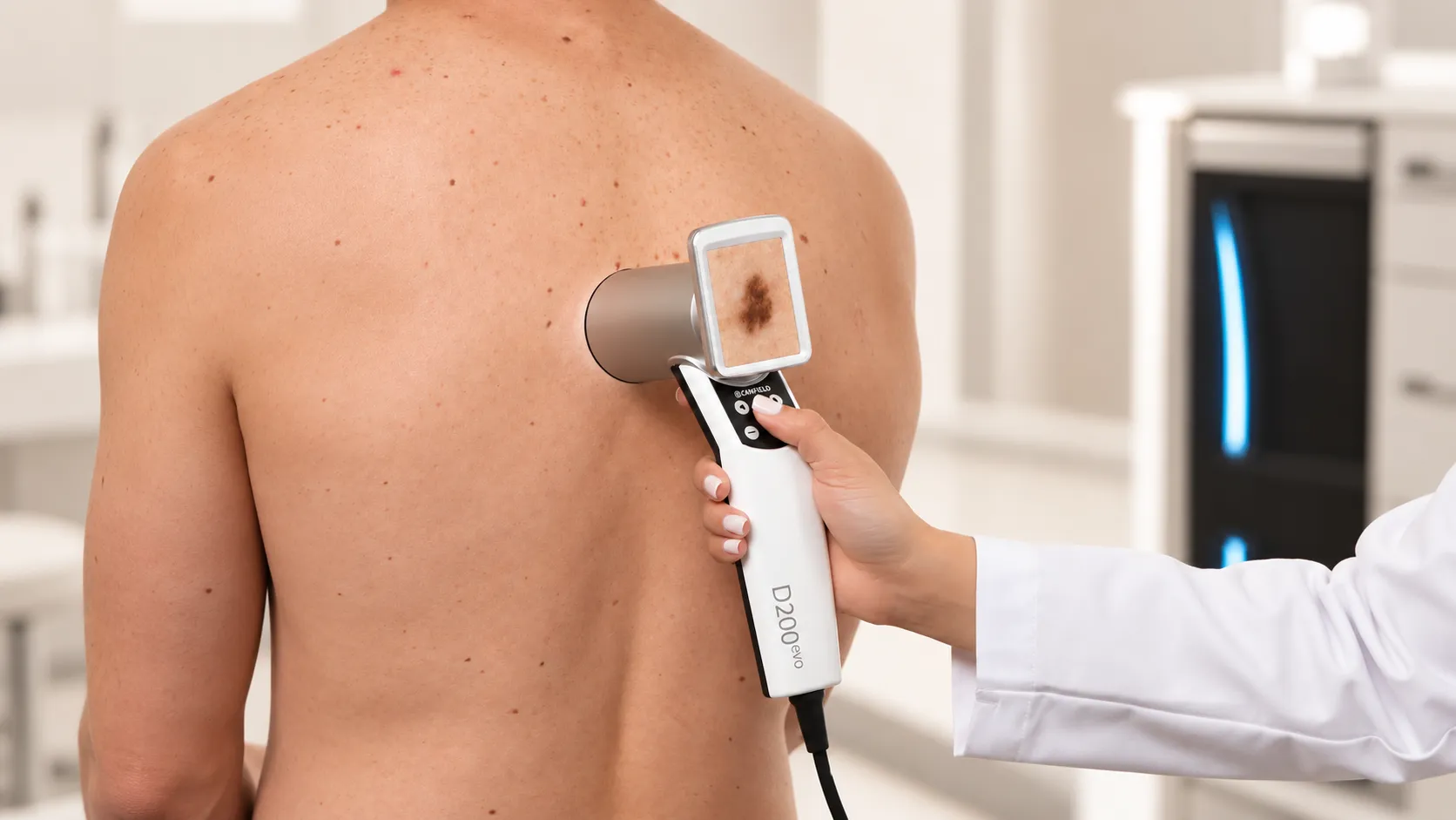
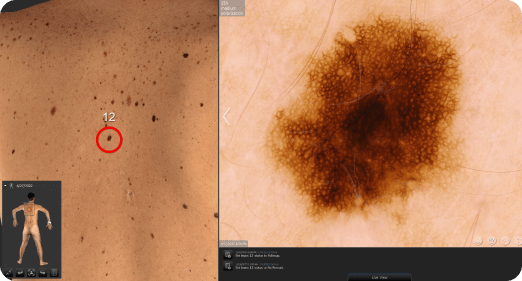
The doctor examines suspicious lesions using a handheld device that magnifies the skin 15 to 200 times. A digital dermatoscope allows for a magnified assessment of the lesion’s structure and pigmentation characteristics, which are not sufficiently visible to the naked eye. Various lighting modes provide additional information for evaluating the lesion. Each close-up image is assigned a unique number.

Benefits

  • Comparability: Standardized photography minimizes differences in lighting and positioning, helping the physician assess changes in skin lesions over time.
  • Traceability: Documented lesions are marked on a skin map so they can be located and compared during future examinations.
  • No ionizing radiation: full-body images are obtained through photography, without skin contact. Depending on the type of examination, dermatoscopy is performed with or without skin contact.
  • We can also see what hasn't changed: a static composition is just as important as a dynamic one.

When is an examination necessary?

Who is this test suitable for?

When the risk is higher

Many moles

A family history of melanoma

A history of melanoma in your own family

Fair skin that burns easily

Sunburns as a child

Use of tanning beds

Irregularly shaped moles

A weakened immune system

When to seek care without waiting

A new mole that appears in adulthood

Change in shape or color

Blurred or irregular border

A growth that is getting bigger

Bleeding, itching, or ulceration

A lesion that looks different from the others

Individual monitoring

After treatment for melanoma or another skin tumor

If there are many atypical moles

If you are undergoing immunosuppressive therapy

Monitor the lesions at intervals specified by your doctor

If removal is necessary, mapping is not a substitute for it

Specialists

Specialists who map birthmarks

BUJ

Frequently Asked Questions

Does it hurt?

The device does not come into contact with you during the examination. The dermatoscope touches the skin lightly.

How long does it take?

The photo session takes about 20 minutes. If it includes a consultation with a dermatologist at the same time, it takes about 50 minutes.

Will I have to undress?

A full-body examination is usually performed while you are in your underwear. The doctor will also discuss with you the examination of areas covered by your underwear and other areas that are difficult to examine. The examination is conducted with respect for your privacy.

Does the machine detect skin cancer on its own?

No. It helps the doctor notice what has changed or appeared for the first time. The doctor makes the diagnosis.

How often should I come back?

The dermatologist determines the timing of the next checkup based on the skin findings and individual risk. A follow-up appointment may be scheduled in 6–12 months or sooner.

Is mapping necessary for everyone?

No. If there are only a few lesions, an examination by a dermatologist and a dermatoscopy may be sufficient. The doctor will assess the need for a full-body screening based on the number of moles, their location, and individual risk.

Who can see my photos?

The images are part of your medical records. Access to them is restricted and is granted in accordance with the clinic’s patient data protection policy.